Errors during cell division are driving the evolution of cancer cells and enabling them to develop resistance to chemotherapy, researchers have established. An eight-year, multi-million-pound collaboration between the University of Dundee and the Max Planck Institute of Molecular Physiology in Germany is now working to unravel the precise molecular mechanisms behind these failures, with the aim of identifying new therapeutic targets.
How cell division errors drive chemotherapy resistance
When cells divide, they must copy their chromosomes and distribute one complete set to each daughter cell. Mistakes in this process lead to aneuploidy — an abnormal number of chromosomes — a condition that is a hallmark of cancer. This chromosomal instability (CIN) not only fuels tumour growth and survival but also drives the emergence of chemotherapy resistance. Research from the Max Planck Institute of Molecular Physiology has previously shown that high rates of chromosome segregation errors in tumour cells directly contribute to both tumour progression and the ability of cancer cells to evade treatment.
The underlying problem lies in the delicate machinery that orchestrates chromosome separation. Improper attachments between chromosomes and the cellular structures that pull them apart create an unstable genome, giving cancer cells the variation they need to adapt and survive under the selective pressure of chemotherapy.
The kinetochore: a molecular machine under the microscope
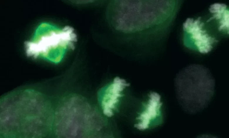
Central to this process is the kinetochore, a complex molecular machine located at the centromere of each chromosome. Its primary role is to ensure accurate chromosome segregation by acting as the attachment point for microtubules — the protein fibres that pull chromosomes to opposite ends of the dividing cell. This attachment is a balancing act: if it is too fragile, chromosomes may be lost; if it is too stable, errors in segregation can become permanent. Kinetochore dysfunction is therefore a key driver of the chromosomal instability seen in many cancers.
The kinetochore’s behaviour is regulated by a system of chemical tags known as phosphates. These phosphate groups attach to proteins, switching them on, and detach to switch them off — a mechanism researchers describe as cellular “light switches”. While scientists have long known which proteins are switched on or off during cell division, the critical missing piece has been the speed at which these switches operate. This rapid on-off cycling, termed phosphorylation–dephosphorylation (PdP) dynamics, has been likened to a “biological morse code” that sends precise instructions to control cell behaviour.
When these signalling pathways malfunction — particularly when the flashing rate of the switches is disrupted — it can trigger uncontrolled cell division and the onset of cancer. Understanding the precise PdP dynamics that govern healthy division is essential to explaining how errors arise in malignant cells and how they might be corrected.
Professor Andrea Musacchio, a director at the Max Planck Institute of Molecular Physiology and a co-investigator on the study, brings specialised expertise in the “biochemical reconstitution of the kinetochore”. This involves rebuilding the kinetochore from its component parts in a test tube, allowing the team to observe its function in isolation. “Our expertise in the biochemical reconstitution of the kinetochore complements the diverse skillsets of our team and gives us the opportunity to understand these patterns during cell division in healthy cells, and what goes wrong in cancer cells that allow them to evolve and become resistant to chemotherapy,” he said.
Decoding the biological morse code
The research is led by Professor Adrian Saurin at the University of Dundee, who is focusing on the “biological morse code” of protein phosphorylation. The collaboration aims to decipher the specific flashing rates of the cellular light switches that are critical for healthy cell division and how their disruption leads to cancer. The work builds on a broader foundation of research across the Max Planck Society. At the Max Planck Institute for Biology of Ageing, scientists have explored links between DNA repair defects, genome instability and cancer, and have developed a compound that targets mitochondria in cancer cells to starve them without severe side effects. Meanwhile, researchers at the Max Planck Institute for Molecular Genetics have elucidated molecular principles of cell division control, finding that checkpoint kinases interact with spindle proteins — a discovery that underscores how incorrect chromosome distribution can lead to cancer.
The study’s ultimate goal is to translate these fundamental insights into new therapeutic strategies. Kinetochore-microtubule attachments, the very mechanism that can go wrong and fuel cancer, are now being evaluated as a compelling target for anti-cancer drugs. Drugs that disrupt the cell cycle, such as CDK4/6 inhibitors, are already in clinical use, and combinations with traditional chemotherapy have shown promise. By understanding exactly how the kinetochore’s “light switches” malfunction in cancer cells, researchers hope to design treatments that can re-establish accurate chromosome segregation and halt the evolution of chemotherapy resistance.