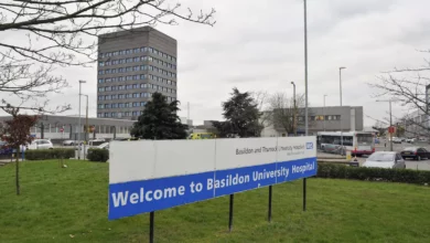
Minimal access surgery offers a durable cure with fewer drawbacks, according to surgeons at Leeds Teaching Hospitals NHS Trust who have performed a UK-first operation to treat a brain aneurysm through the patient’s eye socket. The procedure, carried out on Andrew Wood, a 61-year-old grandfather and builder from Leeds, marks a significant step forward in minimally invasive brain surgery by providing a permanent solution to a potentially fatal condition while eliminating many of the adverse effects associated with traditional open surgery.
Benefits of the keyhole technique
The operation used a keyhole approach to reach the aneurysm directly through the eye socket, avoiding the need for a craniotomy – the removal of a section of the skull. Instead of retracting or cutting through brain tissue to access the swollen blood vessel, the surgical team, led by consultant neurosurgeon Asim Sheikh and consultant maxillofacial surgeon Jiten Parmar, were able to navigate through the natural bony anatomy. The result was a procedure that did not require touching the brain at all, drastically reducing the morbidity traditionally linked to brain surgery.
Advanced preparation was central to the success of the technique. Biomechanical engineers created a bespoke 3D-printed model of Mr Wood’s eye socket, the surrounding skull base and the aneurysm itself. This allowed the team to plan and rehearse the entire operation tailored specifically to his anatomy. Custom 3D-printed retractors were also developed to protect the eye during the surgery, ensuring the delicate structures remained unharmed.
The recovery time was dramatically shorter than with conventional methods. Mr Wood was discharged from hospital after just one night – compared to the typical week-long stay for patients who have undergone a craniotomy. He returned to his work as a builder in May, only weeks after the operation in February. The morning after surgery, he reported no double vision or pain and was able to perform everyday tasks such as making toast and tea, a testament to the minimal disruption to his daily life.
Drawbacks removed by the new approach
Traditional surgical treatment for a brain aneurysm – known as clipping – requires a large incision in the scalp, removal of a portion of the skull, and careful retraction of brain tissue to reach the aneurysm. This open approach carries significant drawbacks: big cuts and scars, extensive incisions on the head, and the inherent risks and recovery challenges of manipulating the brain. The keyhole technique through the eye socket eliminates all of these. There are no large external incisions, no need to open the skull, and no retraction of brain tissue, meaning the common complications of swelling, bleeding, and prolonged recovery are drastically reduced.
The procedure offers the best of both worlds, according to the surgical team – a durable cure with a very low risk of recurrence, comparable to the gold-standard clipping method, while removing nearly all the disadvantages that patients typically face. For Mr Wood, whose aneurysm was discovered incidentally during scans for an unrelated medical issue and who had no symptoms, the minimally invasive option was particularly reassuring. He has said he is proud to have taken part in a pioneering operation and is grateful for the outcome.
Context and significance
This is not the first time the Leeds team has pushed the boundaries of keyhole neurosurgery. In 2024, the same surgeons performed a UK-first operation to remove a brain tumour through a patient’s eye socket, demonstrating the expanding potential of this approach for a range of conditions. The technique joins a growing arsenal of minimally invasive options in UK neurosurgery, including endovascular coiling – where a catheter is guided from the groin to fill the aneurysm with platinum coils – and robotic systems such as the ExcelsiusGPS recently installed at the Royal Victoria Infirmary in Newcastle. 3D printing, virtual reality, artificial intelligence for surgical navigation, and intraoperative MRI are all being integrated to improve precision and outcomes.
For Mr Wood, the journey from diagnosis to recovery has been remarkably swift. He was shocked to learn he had a brain aneurysm, but found the explanation of the eye-socket approach reassuring. His experience underscores what consultant neurosurgeon Asim Sheikh described as a significant step forward: the ability to deliver a durable, permanent cure for a brain aneurysm while cutting down on the major drawbacks of conventional surgery – big incisions, scarring, and the morbidity of going through and retracting the brain. All of that, in this case, was completely taken away.