AI could spot pancreatic cancer warning signs before scans reveal tumours, research indicates
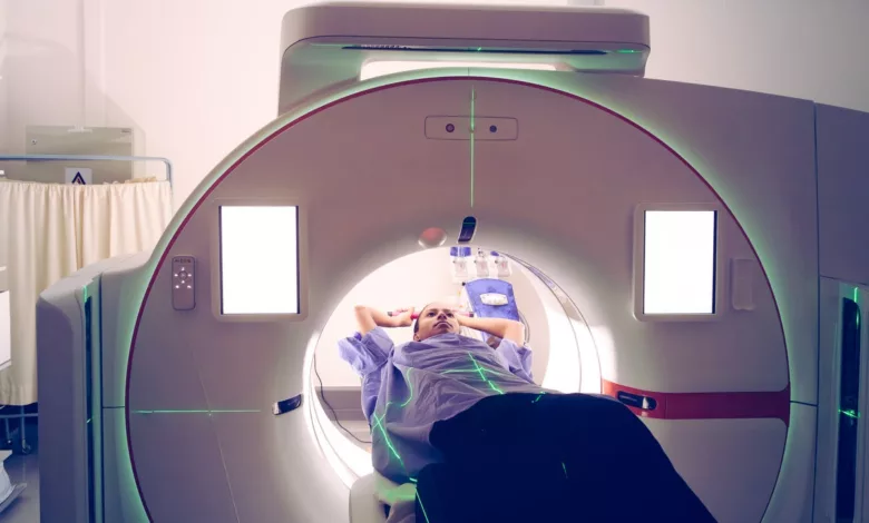
A new artificial intelligence model has demonstrated the ability to spot signs of pancreatic cancer on CT scans up to three years before a clinical diagnosis, according to a study published in the journal Gut.
Developed by researchers at the Mayo Clinic, the AI framework — named REDMOD, short for Radiomics-based Early Detection MODel — identifies “subvisual” patterns in the pancreas that are invisible to the human eye. The technology represents a potential breakthrough in a disease where the five-year survival rate remains just 13 percent, and where more than 85 percent of patients are diagnosed only after the cancer has already spread.
Pancreatic tumours are notoriously difficult to catch early. The organ sits deep within the abdomen, making physical examinations ineffective, and there is no routine screening available for the general public. By the time symptoms such as abdominal pain or weight loss emerge, the cancer has typically reached an advanced stage. Projections show it is set to become the second-leading cause of cancer-related death in the United States by 2030.
How the AI detects the invisible
Unlike radiologists, who are trained to look for a measurable mass, the AI model scans for far subtler signs. It analyses “radiomic” features — minute textural disruptions in the pancreatic tissue that would be imperceptible even to a trained eye. The model's predictive power comes principally from multi-scale wavelet-filtered textural features that capture what the researchers describe as subvisual architectural disruptions.
Dr. Ajit Goenka, a Mayo Clinic radiologist and senior author of the study, explained that the AI can identify the signature of abnormal cells that protect the cancer from the immune system — a phenomenon scientists have long recognised at a molecular level but have struggled to visualise on standard imaging. “We knew, based on the biology of the disease, that this is not something which is coming all of a sudden in three months,” he said. “We knew that the signal was there. We just needed to find a way to be able to detect it.”
REDMOD also includes an automated pancreatic segmentation tool that clearly delineates the pancreas from surrounding tissues and organs. This removes the need for manual segmentation, which can be variable in accuracy. The AI analysis further revealed that patients who later developed pancreatic cancer had significantly smaller pancreatic volumes on their CT scans compared to controls, a finding that may itself serve as an early indicator.
Performance against human radiologists
The model was trained on nearly 1,000 CT scans taken from patients who were originally screened for unrelated conditions but went on to develop pancreatic cancer. In total, around 2,000 scans were analysed across the training and validation phases. On an independent test set of 493 scans, REDMOD identified 73 percent of pre-diagnostic cancers at a median of approximately 16 months before clinical diagnosis. This detection rate was nearly double the 39 percent sensitivity achieved by board-certified radiologists reviewing the same scans without AI assistance.
For scans taken more than two years before a formal diagnosis, the gap widened further: the AI achieved 68 percent sensitivity against 23 percent for the human doctors — nearly three times as effective. The model recorded an Area Under the Curve (AUC) of 0.82, a measure of its overall diagnostic accuracy. It also demonstrated strong longitudinal stability, producing consistent results across multiple scans from the same patient over time, and generalisable specificity across CT scans from different institutions and imaging systems.
Dr. Daniel Jeong, a radiologist at Moffitt Cancer Center who was not involved in the research, described the limitations of current practice. “I analyze these images every day,” he said. “We’re really looking for a measurable mass that could represent the cancer. So these tumors need to grow to a certain level to become visible.”
Limitations and the road ahead
Despite the promising results, the researchers caution that the tool is not yet ready for routine clinical use. The model is currently being evaluated in a clinical trial that requires three to five years of patient monitoring to confirm its accuracy in real-time. Mayo Clinic is advancing this work through a programme called Artificial Intelligence for Pancreatic Cancer Early Detection (AI-PACED).
The study has acknowledged limitations, including a lack of ethnic diversity within the cohort, which will need to be addressed before the model can be applied more broadly. Nevertheless, researchers believe the technology could eventually serve as a triage tool for individuals at high risk, such as those with a family history of the disease or new-onset diabetes — a group that is the focus of the UK-Early Detection Initiative for Pancreatic Cancer (UK-EDI), which targets those aged 50 and over newly diagnosed with diabetes.
In the UK, pancreatic cancer is the fifth most common cause of cancer death, accounting for around 6 percent of all cancer fatalities. Five-year survival rates hover at about 7.3 percent, and as low as 4.3 percent for ten-year survival. Key modifiable risk factors in the UK include smoking, which accounts for around 20 to 22 percent of cases, and being overweight or obese, which contributes to over 10 to 12 percent.
The research was supported in part by the Hoveida Family Foundation and joins a wave of recent advancements in the field, including early-stage trials for mRNA vaccines, experimental drugs, and new blood and breath tests for early detection. “In a disease where we have been just wandering in darkness for decades, this is a milestone that shows us the finish line, but we still have to get to the finish line,” Goenka said.